LA DERMATOSCOPIA
La dermatoscopia, también conocida como dermatoscopia, microscopía de epiluminiscencia o microscopía de superficie, es una técnica de diagnóstico no invasiva que permite la observación en vivo con aumento a través de un lente de lesiones y estructuras cutáneas que no son visibles a simple vista.
Esta técnica utiliza un instrumento portátil llamado dermatoscopio, que está equipado con una fuente de luz transiluminadora y una lente de aumento, que normalmente proporciona un aumento de alrededor de 10x.
ORÍGENES DE LA DERMATOSCOPIA:
La dermatoscopia, también conocida como microscopía de epiluminiscencia, comenzó a desarrollarse en la década de 1990. Sin embargo, sus raíces se remontan a técnicas anteriores de examen de la piel que utilizaban métodos ópticos, remontándose a la década de los 60 - 70, pero fue en los 90 cuando adquirió notoriedad.
La aplicación primordial de la dermatoscopia es la evaluación de lesiones cutáneas pigmentadas para mejorar la precisión diagnóstica de afecciones, como el melanoma y el carcinoma basocelular. Al visualizar las estructuras subsuperficiales, los médicos pueden identificar patrones y colores específicos que son indicativos de malignidad, lo que ayuda a diferenciar entre lesiones malignas y benignas.
Además de su uso en oncología, la dermatoscopia se ha ampliado para incluir el diagnóstico de diversas afecciones dermatológicas, como enfermedades inflamatorias (inflamoscopia), trastornos del cabello y el cuero cabelludo (tricoscopia) y anomalías de las uñas (onicoscopia). La técnica también se puede aplicar a lesiones mucosas, lo que proporciona una alternativa no invasiva a las biopsias en áreas de difícil acceso o propensas a sangrar.
Existen dos técnicas principales para realizar dermatoscopia: de contacto y sin contacto. En el método de contacto, el dermatoscopio se aplica directamente a la piel con una interfaz líquida, (aceite de cedro o alcohol), mientras que el método sin contacto no requiere contacto directo con la piel.
Las formas avanzadas de dermatoscopia, como la videodermatoscopia, ofrecen la ventaja de además de proporcionar aumento, hay una capacidad digital para el almacenamiento y análisis de imágenes, lo cual es útil para el posterior seguimiento de las lesiones.
La tecnología ha avanzado tanto que las cámaras de los móviles (Smartphones) o celulares, hoy día ofrecen una opción en la cámara tipo MACRO, EL cual proporciona un aumento comparativo a un dermatoscopio DIGITAL y el costo es mucho menor.
De hecho EXISTE una técnica donde al lente del móvil se le sobrepone la lente de las lectoras de DVD y el aumento es igual o SUPERIOR a un dermatoscopio DIGITAL y la GRAN VENTAJA sobre un dermatoscopia convencional es que puedes ver la LESIÓN EN toda la pantalla del móvil, pudiendo tomar foto de la misma o video, es decir, el móvil funciona como VIDEO DERMATOSCOPIO.
Esto nos permite clasificar los dermatoscopios en:
1.) Dermatoscopios analogicos: los cuales pueden tener o no contacto con la piel con luz o sin luz.
Dematoscopio Analogico
2.) Dermatoscopios digitales: Equipados con una cámara de video que puede tomar instantáneas, grabar las lesiones, y almacenarlas, útil para el seguimiento de las lesiones que pudieran tener una tendencia a malignizarse.
3.) Dermatoscopios en Smartphones: como les comente, existen en el mercado móviles con cámaras que tiene un lente MACRO muy potente, los cuales cumplen la misma función que un dermatoscopio DIGITAL y son mucho más económicos.
Dermatoscopio-Smartphone
USOS DE LA DERMATOSCOPIA:
1.) Detección Temprana del Melanoma: Uno de los usos más importantes es el diagnóstico precoz del melanoma y otros tipos de cáncer de piel. La técnica permite identificar características específicas en las lesiones que podrían indicar malignidad.
2.) Evaluación de Lesiones Benignas: Además del melanoma, se utiliza para evaluar lunares benignos, carcinomas basocelulares y otros tumores cutáneos.
3.) Diagnóstico de Enfermedades Infecciosas: La dermatoscopia puede ayudar en el diagnóstico de condiciones como sarna o piojos al permitir visualizar estructuras que no son evidentes a simple vista.
4.) Trastornos del Cabello y Uñas: También es útil para diagnosticar problemas relacionados con el cabello y las uñas, como alopecia areata o infecciones fúngicas.
5.) Dermatoscopia digital: Permite realizar un seguimiento continuo de las lesiones cutáneas sospechosas, almacenando imágenes para comparaciones futuras y ayudando a detectar cambios significativos con el tiempo. El dermatoscopio convencional no ofrece esta opción, la alternativa es la explicada anteriormente, utilizando la lente de un móvil modificada, o un Smartphone con un lente MACRO de gran capacidad de aumento.
En resumen, la dermatoscopia es una herramienta valiosa en dermatología que mejora la sensibilidad y especificidad del diagnóstico, disminuye la necesidad de procedimientos invasivos y ayuda en el seguimiento de las respuestas al tratamiento.
Saludos,,,
Dr. José Lapenta.
ENGLISH
Dermatoscopy, also known as dermatoscopy, epiluminescence microscopy, or surface microscopy, is a noninvasive diagnostic technique that allows live observation with magnification through a lens of skin lesions and structures that are not visible to the naked eye.
This technique uses a portable instrument called a dermatoscope, which is equipped with a transilluminating light source and a magnifying lens, typically providing a magnification of around 10x.
ORIGINS OF DERMATOSCOPY:
Dermatoscopy, also known as epiluminescence microscopy, began to develop in the 1990s. However, its roots go back to earlier skin examination techniques that used optical methods, dating back to the 1960s - 1970s, but it was in the 1990s that it gained notoriety.
The primary application of dermoscopy is the evaluation of pigmented skin lesions to improve diagnostic accuracy for conditions such as melanoma and basal cell carcinoma. By visualizing subsurface structures, physicians can identify specific patterns and colors that are indicative of malignancy, helping to differentiate between malignant and benign lesions.
In addition to its use in oncology, dermoscopy has been expanded to include the diagnosis of a variety of dermatologic conditions, such as inflammatory diseases (inflammoscopy), hair and scalp disorders (trichoscopy), and nail abnormalities (onychoscopy). The technique can also be applied to mucosal lesions, providing a noninvasive alternative to biopsies in areas that are difficult to access or prone to bleeding.
There are two main techniques for performing dermoscopy: contact and noncontact. In the contact method, the dermatoscope is applied directly to the skin with a liquid interface, (cedar oil or alcohol), while the non contact method does not require direct contact with the skin.
Advanced forms of dermatoscopy, such as video dermatoscopy, offer the advantage of providing magnification as well as digital image storage and analysis, which is useful for subsequent follow-up of lesions.
Technology has advanced so much that mobile phone cameras (Smartphones) or cell phones, today offer an option in the MACRO type camera, which provides a magnification similar to a DIGITAL dermatoscope and the cost is much lower.
In fact, there is a technique where the lens of DVD players is superimposed on the mobile phone lens and the magnification is equal to or GREATER than a DIGITAL dermatoscope and the GREAT ADVANTAGE over a conventional dermatoscopy is that you can see the LESION ON the entire mobile phone screen, being able to take a photo or video of it, that is, the mobile phone works as a VIDEO DERMATOSCOPY.
This allows us to classify dermatoscopes into:
1.) Analog dermatoscopes: which may or may not have contact with the skin with or without light.
Analog Dermatoscopy
2.) Digital dermatoscopes: Equipped with a video camera that can take snapshots, record the lesions, and store them, useful for monitoring lesions that may have a tendency to become malignant.
Digital Dermatoscopy
3.) Dermatoscopes on Smartphones: as I mentioned, there are mobile phones on the market with cameras that have a very powerful MACRO lens, which perform the same function as a DIGITAL dermatoscope and are much cheaper.
Smartphone-Dermatoscopy
USES OF DERMATOSCOPY:
1.) Early Detection of Melanoma: One of the most important uses is the early diagnosis of melanoma and other types of skin cancer. The technique allows for the identification of specific characteristics in lesions that could indicate malignancy.
2.) Evaluation of Benign Lesions: In addition to melanoma, it is used to evaluate benign moles, basal cell carcinomas, and other skin tumors.
3.) Diagnosis of Infectious Diseases: Dermoscopy can help in the diagnosis of conditions such as scabies or lice by allowing the visualization of structures that are not evident to the naked eye.
4.) Hair and Nail Disorders: It is also useful in diagnosing hair and nail-related problems, such as alopecia areata or fungal infections.
5.) Digital Dermoscopy: It allows continuous monitoring of suspicious skin lesions, storing images for future comparison and helping to detect significant changes over time. The conventional dermatoscope does not offer this option, the alternative is the one explained above, using the lens of a modified mobile phone, or a Smartphone with a high magnification MACRO lens.
In summary, dermoscopy is a valuable tool in dermatology that improves the sensitivity and specificity of diagnosis, decreases the need for invasive procedures, and helps in monitoring treatment responses.
Greetings...
Dr. José Lapenta R.
EDITORIAL ESPANOL:
====================
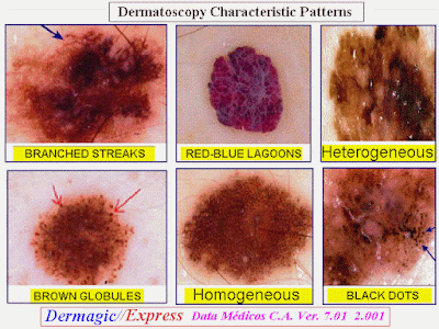
Hola Amigos Dermagicos, El tema de hoy LA DERMATOSCOPIA. Método mediante el cual se visualizan una serie de características en las lesiones pigmentadas de la piel, que van más allá del clásico ABCD. Estas 15 referencias nos hablan de los beneficios de este procedimiento y en el attach 3 láminas ilustrativas del tema.
Próximas ediciones: * Los hongos y sus familias
* Los Eumicetos
Saludos,,,
Dr. José Lapenta R.,,,
EDITORIAL ENGLISH:
===================
Hello Dermagics friends, today's topic THE DERMATOSCOPY. Method by means of which you can visualize a series of characteristic in the pigmented skin lesions that explain more than the classic ABCD. These 15 references speak about of the benefits of this procedure and in the attach 3 illustrative sheets of the topic.
Next editions: * The fungus and their families
* The Eumycetos
Greetings,,,
Dr. José Lapenta R.
======================================================================
DERMAGIC/EXPRESS(41)
======================================================================
LA DERMATOSCOPIA/ DERMATOSCOPY
======================================================================
1.) DERMATOSCOPY
2.) Systematic review of the diagnostic accuracy of dermatoscopy in detecting malignant melanoma.
3.) [Dermatoscopy. An investigative method for the diagnosis of pigmented skin tumors]
4.) Dermatoscopy: usefulness in the differential diagnosis of cutaneous pigmentary lesions.
5.) The ABCD rule of dermatoscopy. High prospective value in the diagnosis of doubtful melanocytic skin lesions.
6.) [The ABCD rule in dermatoscopy: analysis of 500 melanocytic lesions]
7.) Early diagnosis of melanoma by surface microscopy (dermatoscopy).
8.) [Improving clinical diagnosis of pigmented skin changes in childhood with the dermatoscope]
9.) Frequency and morphologic characteristics of invasive melanomas lacking specific surface microscopic features.
10.) A sensitivity and specificity analysis of the surface microscopy features of invasive melanoma.
11.) Distinctive dermatoscopic features of acral lentiginous melanoma in situ from plantar melanocytic nevi and their histopathologic correlation.
12.) [Classification, diagnosis and differential diagnosis of malignant melanoma]
13.) 'Dimpling' is not unique to dermatofibromas.
14.) Epiluminescence Microscopy for the Diagnosis of Doubtful Melanocytic Skin Lesions
Comparison of the ABCD Rule of Dermatoscopy and a New 7-Point Checklist Based on Pattern Analysis
15.) Dermatoscopy Transforms Invisible to Visible in Pigmented Lesions.
16.) Dermatoscopy: Alternative Uses in Daily Clinical Practice.
17.) Dermoscopy for the Family Physician.
19.) Two Controversies Confronting Dermoscopy or Dermatoscopy: Nomenclature and Results.
20.) Dermoscopy of Oral and Genital Mucosal Lesions: A Descriptive Cross-Sectional Study Protocol.
21.) Clinical Techniques in Veterinary Dermatology: Dermoscopy.
========================================================================
========================================================================
1.) DERMATOSCOPY
========================================================================
In vivo cutaneous surface microscopy, EpiLuminescence microscopy (ELM), dermoscopy, dermatoscopy and magnified oil immersiondiascopy, are terms that describe the use of an incident light magnification system to examinecutaneous lesions, that are not perceptible by thedermatologist during his clinical observation. The examinationcomes made supporting on the skin previously cover with a thinlayer of oil by immersion the objective of a simple microscope or compound. Alsouse a tool to optic fibers that it allows of visualize on a protectionthe image (videodermatoscopiy) .The image can also be digitalized and processed from softwareand sent in Local Network Area (LAN),Wide AreaNetwork (WAN), INTERNET, INTRANET (Teledermatoscopy) . The goal of the dermatoscopy is the precocious and accurate diagnosis of the melanoma in his initial phases, for the diagnosis of melanocyticlesions and excludes non-melanocytic lesions and between benign and malignant lesions with a sensibilitydiagnostic of the 90% .The dermatoscopy betters the accuracy of the diagnosis of melanoma in theclinical practice[] .One studies however it has shown a meaningful diminution in sensibility when it is practiced from dermathologyst without experience.
Copyright © 1997 [Stefano Lorenzi].
========================================================================
2.) Systematic review of the diagnostic accuracy of dermatoscopy in detecting malignant melanoma.
========================================================================
Author
Mayer J
Address
Danila Dilba Medical Service, Darwin, NT. jmayer@racgp.org.au
Source
Med J Aust, 167(4):206-10 1997 Aug 18
Abstract
OBJECTIVE: To assess the evidence that dermatoscopy improves the accuracy of diagnosis of melanomas in clinical practice. DATA SOURCES: MEDLINE 1983-January 1997, EMBASE 1980-1996, and bibliographies of retrieved articles. STUDY SELECTION AND DATA EXTRACTION: Studies selected were original studies with formal methods and results sections comparing diagnostic accuracy of dermatoscopy for malignant melanoma with another clinical method; the criterion standard was excision biopsy with histopathological examination; and accuracy of dermatoscopic diagnosis was determined over a spectrum of stages of melanoma and skin lesions commonly confused with melanoma. Data were extracted by a single observer. DATA SYNTHESIS: 579 articles were identified; six studies met the inclusion criteria. Positive likelihood ratios for dermatoscopy for diagnosis of melanoma ranged from 2.9 to 10.3. Dermatoscopy had 10%-27% higher sensitivity than clinical diagnosis in the two studies with the most clinically equivocal lesions. However, when sensitivity of clinical diagnosis was more than 84%, sensitivity of dermatoscopy was only slightly higher. One study of dermatologists with no training in dermatoscopy showed a significant decrease in sensitivity. CONCLUSIONS: Variability between studies in methods, observers and types of pigmented skin lesions and lack of studies in primary care make generalisation of results difficult. Dermatoscopy appeared not to improve the accuracy of diagnosis enough to alter the clinical management of most pigmented skin lesions. Further research with more explicit methods is needed.
Language
========================================================================
3.) [Dermatoscopy. An investigative method for the diagnosis of pigmented skin tumors]
========================================================================
Author
Weismann K; Osterlind AL; Thomsen HK
Address
Dermato-venerologisk afdeling A, Bispebjerg Hospital, København.
Source
Ugeskr Laeger, 157(2):147-52 1995 Jan 9
Abstract
Dermatoscopy is a non-invasive investigative technique which makes it possible to evaluate the pigmented structures of the epidermis, the dermo-epidermal junction and the papillary dermal layer. The melanin pigmentation of the epidermal basal cell layers, the so-called pigment network is the primary target of dermatoscopy. A number of diagnostic criteria and variables for the dermatoscopy of pigmented skin lesions have been developed. Cutaneous malignant melanoma is still increasing in incidence and mortality. An early diagnosis is decisive for the prognosis. Therefore, it is urgent to develop practical investigative methods which can increase the diagnostic sensitivity. Dermatoscopy is such a method, but it demands training and experience. Lack of suspicious findings by dermatoscopy does not exclude malignancy. A safe diagnosis can only be obtained by a histological examination.
========================================================================
4.) Dermatoscopy: usefulness in the differential diagnosis of cutaneous pigmentary lesions.
========================================================================
Author
Cristofolini M; Zumiani G; Bauer P; Cristofolini P; Boi S; Micciolo R
Address
Department of Dermatology, S. Chiara Hospital, Trento, Italy.
Source
Melanoma Res, 4(6):391-4 1994 Dec
Abstract
Dermatoscopy has been reported to give valid information in the differential diagnosis of cutaneous pigmentary lesions. Using a dermatoscopy Delta 10 Heine optotechnique, we evaluated 220 pigmented lesions during a health campaign for the early diagnosis of cutaneous melanoma and compared clinical and dermatoscopic diagnosis. Histologic diagnosis was carried out after removal of the lesions. Sensitivity, specificity, positive predictive value, negative predictive value and overall agreement were evaluated. In our experience clinical and dermatoscopic diagnosis gave similar results; sensitivity and specificity were slightly better for dermatoscopy than for clinical diagnosis. The agreement between clinical and dermatoscopic diagnosis was better in histologically negative lesions. Dermatoscopy is useful in the diagnosis of pigmentary lesions, but clinical diagnosis by experienced dermatological staff, is unreplaceable, especially during a health campaign for the early diagnosis of cutaneous melanoma.
Language
========================================================================
5.) The ABCD rule of dermatoscopy. High prospective value in the diagnosis of doubtful melanocytic skin lesions.
========================================================================
Author
Nachbar F; Stolz W; Merkle T; Cognetta AB; Vogt T; Landthaler M; Bilek P; Braun-Falco O; Plewig G
Address
Department of Dermatology, University of Munich, Germany.
Source
J Am Acad Dermatol, 30(4):551-9 1994 Apr
Abstract
BACKGROUND: The difficulties in accurately assessing pigmented skin lesions are ever present in practice. The recently described ABCD rule of dermatoscopy (skin surface microscopy at x10 magnification), based on the criteria asymmetry (A), border (B), color (C), and differential structure (D), improved diagnostic accuracy when applied retrospectively to clinical slides. OBJECTIVE: A study was designed to evaluate the prospective value of the ABCD rule of dermatoscopy in melanocytic lesions. METHODS: In 172 melanocytic pigmented skin lesions, the criteria of the ABCD rule of dermatoscopy were analyzed with a semiquantitative scoring system before excision. RESULTS: According to the retrospectively determined threshold, tumors with a score higher than 5.45 (64/69 melanomas [92.8%]) were classified as malignant, whereas lesions with a lower score were considered as benign (93/103 melanocytic nevi [90.3%]). Negative predictive value for melanoma (True-Negative divided by [True-Negative+False-Negative]) was 95.8%, whereas positive predictive value (True-Positive divided by [True-Positive+False-Positive]) was 85.3%. Diagnostic accuracy for melanoma (True-Positive divided by [True-Positive+False-Positive+False-Negative]) was 80.0%, compared with 64.4% by the naked eye. Melanoma showed a mean final dermatoscopy score of 6.79 (SD, +/- 0.92), significantly differing from melanocytic nevi (mean score, 4.27 +/- 0.99; p < 0.01, U test). CONCLUSION: The ABCD rule can be easily learned and rapidly calculated, and has proven to be reliable. It should be routinely applied to all equivocal pigmented skin lesions to reach a more objective and reproducible diagnosis and to obtain this assessment preoperatively.
========================================================================
6.) [The ABCD rule in dermatoscopy: analysis of 500 melanocytic lesions]
========================================================================
Author
Feldmann R; Fellenz C; Gschnait F
Address
Dermatologische Abteilung, Krankenhaus der Stadt Wien-Lainz.
Source
Hautarzt, 49(6):473-6 1998 Jun
Abstract
500 melanocytic lesions were examined by dermatoscopy using the ABCD rule prior to excision and histologic diagnosis. Regular nevi (n = 272) exhibited a score of 3.55 +/- 0.87, nevi with histologic signs of dysplasia (n = 190) a score of 4.0 +/- 0.68 and melanomas (n = 30) a score of 5.08 +/- 1.24. This study suggests that the ABCD rule of dermatoscopy greatly facilitates the evaluation of melanocytic lesions. When the dermatoscopic score is higher than 4.2, melanoma should be considered.
========================================================================
7.) Early diagnosis of melanoma by surface microscopy (dermatoscopy).
========================================================================
Author
Paschoal FM
Address
Department of Dermatology, Escola Paulista de Medicina-S~ao Paulo, Brazil.
Source
Rev Paul Med, 114(4):1220-1 1996 Jul-Aug
Abstract
The main objective of surface microscopy is the early and accurate diagnosis of melanoma in its initial phases of evolution and infiltration. Since the development of the dermatoscope in the 1990's, surface microscopy has become a simple technique. Differential diagnosis of pigmented skin lesions can be achieved with a diagnostic sensitivity of about 90 percent, and the proper differentiation of pigmented melanocytic and non-melanocytic lesions, and malignant and benign melanocytic lesions, may also be safely determined.
========================================================================
8.) [Improving clinical diagnosis of pigmented skin changes in childhood with the dermatoscope]
========================================================================
Author
Stolz W; Bilek P; Merkle T; Landthaler M; Braun-Falco O
Address
Dermatologische Klinik und Poliklinik, Ludwig-Maximilians-Universit¨at, M¨unchen.
Source
Monatsschr Kinderheilkd, 139(2):110-3 1991 Feb
Abstract
Prompted by the development of the lightweight, inexpensive and simple to use dermatoscopy, skin surface microscopy can now be applied in daily practice. The case reports presented here document the diagnostic improvement achieved with dermatoscopy in pigment skin lesions of children.
========================================================================
9.) Frequency and morphologic characteristics of invasive melanomas lacking specific surface microscopic features.
========================================================================
Author
Menzies SW; Ingvar C; Crotty KA; McCarthy WH
Address
Department of Surgery (Sydney Melanoma Unit), University of Sydney, Australia.
Source
Arch Dermatol, 132(10):1178-82 1996 Oct
Abstract
OBJECTIVES: To create a simple diagnostic method for invasive melanoma with in vivo cutaneous surface microscopy (epiluminescence microscopy, dermoscopy, dermatoscopy) and to analyze the incidence and characteristics of those invasive melanomas that had no diagnostic features by means of hand-held surface microscopes. DESIGN: Pigmented skin lesions were photographed in vivo with the use of immersion oil. All were excised and reviewed for histological diagnosis. A training set of 62 invasive melanomas and 159 atypical nonmelanomas and a test set of 45 invasive melanomas and 119 atypical non-melanomas were used. Images from the training set were scored for 72 surface microscopic features. Those features with a low sensitivity (0%) and high specificity (> 85%) were used to create a simple diagnostic model for invasive melanoma. SETTING: All patients were recruited from the Sydney (Australia) Melanoma Unit (a primary case and referral center). PATIENTS: A random sample of patients whose lesions were excised, selected from a larger database. MAIN OUTCOME MEASURES: Sensitivity and specificity of the model for diagnosis of invasive melanona. RESULTS: The model gave a sensitivity of 92% (98/107) and specificity of 71%. Of the 9 "featureless" melanomas the model failed to detect, 6 were pigmented and thin and had a pigment network. The other 3 were thicker, hypomelanotic lesions lacking a pigment network, some with prominent telangiectases, and all with only small areas of pigment. All featureless melanomas noted by the patients had a history of change in color, shape, or size. CONCLUSIONS: Surface microscopy does not allow 100% sensitivity in diagnosing invasive melanoma and therefore cannot be used as the sole indicator for excision. Clinical history is an important consideration when featureless lesions are diagnosed.
========================================================================
10.) A sensitivity and specificity analysis of the surface microscopy features of invasive melanoma.
========================================================================
Author
Menzies SW; Ingvar C; McCarthy WH
Address
Sydney Melanoma Unit, Department of Surgery, University of Sydney, Australia.
Source
Melanoma Res, 6(1):55-62 1996 Feb
Abstract
In vivo cutaneous surface microscopy, epiluminescence microscopy, dermoscopy, dermatoscopy and magnified oil immersion diascopy, are terms that describe the use of an incident light magnification system to examine cutaneous lesions, usually with immersion oil at the skin-microscope interface. The result is the visualization of a multitude of morphological features, not visible with the naked eye, that enhance the clinical diagnosis of nearly all pigmented lesions. Sixty-two invasive melanomas and 159 randomly selected non-melanoma pigmented lesions were used in the study. The non-melanomas, while randomly selected from a large data base, were all clinically atypical. Using the x 10 magnification of hand-held surface microscopes (Dermatoscope, Episcope), we present an analysis of 72 surface microscopic variables (constituting over 15,000 single observations) for the diagnosis of invasive melanoma. Forty of the 72 features studied were shown to differ significantly between invasive melanoma and non-melanoma pigmented lesions. Blue-white veil, multiple brown dots, radial streaming and pseudopods had a specificity greater than 95% for melanoma. Two features, symmetrically irregular pigment (non-uniform pigmentation with point and axial symmetry) and the presence of a single colour, had a sensitivity of 0%, i.e. were absent, in melanoma. The other significant features are presented, with their sensitivity and specificity for melanoma.
========================================================================
11.) Distinctive dermatoscopic features of acral lentiginous melanoma in situ from plantar melanocytic nevi and their histopathologic correlation.
========================================================================
Author
Kawabata Y; Tamaki K
Address
University of Tokyo, Faculty of Medicine, Department of Dermatology, Tokyo, Japan.
Source
J Cutan Med Surg, 2(4):199-204 1998 Apr
Abstract
BACKGROUND: An acral lentiginous melanoma in situ on the sole is often difficult to differentiate with the naked eye from an acquired plantar melanocytic nevus. Recent technical advances in epiluminescence microscopy have contributed to the differentiation of these two pigmented skin lesions. OBJECTIVE: In this study, the correlation between dermatoscopic and histopathologic findings of acral lentiginous melanoma in situ on the sole are compared to those of acquired plantar melanocytic nevi. METHODS: Three acral lentiginous melanomas in situ on the sole, and two cases of acral lentiginous melanoma were compared with 50 acquired plantar melanocytic nevi by means of dermatoscopy and histopathology. Results: The dermatoscopic surface profiles of acquired melanocytic nevi were composed of linear pigmentation accentuated mainly on the sulcus superficialis. Histologically, some areas of the sulcus superficialis corresponded to rete ridges of the epidermis, and nests of nevus cells were also often located there. In contrast, the acral lentiginous melanomas in situ showed diffuse, irregularly shaped pigmentation distributed in a disorderly fashion over the entire surface. Histologically, isolated areas of proliferation and small nest formations of atypical melanocytes were irregularly distributed in the epidermis. CONCLUSION: A distinctive dermatoscopic feature of acral lentiginous melanoma in situ is diffuse and irregular pigmentation over the entire surface of the lesion. This feature is helpful for differentiating acral lentiginous melanoma in situ from acquired plantar melanocytic nevi.
========================================================================
12.) [Classification, diagnosis and differential diagnosis of malignant melanoma]
========================================================================
Author
Stolz W; Landthaler M
Address
Klinik und Poliklinik f¨ur Dermatologie, Universit¨at Regensburg.
Source
Chirurg, 65(3):145-52 1994 Mar
Abstract
Due to the recent increase of incidence of malignant melanoma and due to the significance of early detection for a definite cure from the disease, diagnosis and differential diagnosis of malignant melanoma are very important. At first glance the malignant potential of a pigmented mole can be evaluated by the macroscopical ABCD rule (Asymmetry, irregular Border, different Colors, and Diameter larger than 6 mm). In addition, also the history of the patient might be helpful. Thus a malignant melanoma should be considered when a patient reports a new rapidly growing pigmented lesion or a change in an existing mole in color, size, shape, and surface. Itching or burning should also arouse the suspicion of malignant change. Risk factors for the development of a malignant melanmoma are a high number of benign melanocytic nevi, large congenital melanocytic nevi, fair skin types with a tendency to sunburns and a malignant melanoma in the family of the patient. With dermatoscopy, which is skin surface microscopy at 10x magnification, the difficult macroscopical differential diagnosis is facilitated, because this technique opens a new dimension between macroscopy and microscopy.
========================================================================
13.) 'Dimpling' is not unique to dermatofibromas.
========================================================================
Author
Meffert JJ; Peake MF; Wilde JL
Address
Department of Dermatology, Brooke Army Medical Center, Fort Sam Houston, Tex, USA.
Source
Dermatology, 195(4):384-6 1997
Abstract
The dimpling of the skin with lateral compression or 'Fitzpatrick's sign' is considered by many to be pathognomonic for dermatofibromas (DFs). Despite the description of this sign in all major textbooks, not all DFs dimple and all that dimple are not DFs. Other diagnostic investigations such as the use of dermatoscopy may help to confirm the clinical suspicion of DF.
========================================================================
14.) Epiluminescence Microscopy for the Diagnosis of Doubtful Melanocytic Skin Lesions
Comparison of the ABCD Rule of Dermatoscopy and a New 7-Point Checklist Based on Pattern Analysis
========================================================================
Giuseppe Argenziano, MD; Gabriella Fabbrocini, MD; Paolo Carli, MD; Vincenzo De Giorgi, MD; Elena Sammarco, MD; Mario Delfino, MD
Objective: To compare the reliability of a new 7-point checklist based on simplified epiluminescence microscopy (ELM) pattern analysis with the ABCD rule of dermatoscopy and standard pattern analysis for the diagnosis of clinically doubtful melanocytic skin lesions.
Design: In a blind study, ELM images of 342 histologically proven melanocytic skin lesions were evaluated for the presence of 7 standard criteria that we called the "ELM 7-point checklist." For each lesion, "overall" and "ABCD scored" diagnoses were recorded. From a training set of 57 melanomas and 139 atypical nonmelanomas, odds ratios were calculated to create a simple diagnostic model based on identification of major and minor criteria for the "7-point scored" diagnosis. A test set of 60 melanomas and 86 atypical nonmelanomas was used for model validation and was then presented to 2 less experienced ELM observers, who recorded the ABCD and 7-point scored diagnoses.
Settings: University medical centers.
Patients: A sample of patients with excised melanocytic lesions.
Main Outcome Measures: Sensitivity, specificity, and accuracy of the models for diagnosing melanoma.
Results: From the total combined sets, the 7-point checklist gave a sensitivity of 95% and a specificity of 75% compared with 85% sensitivity and 66% specificity using the ABCD rule and 91% sensitivity and 90% specificity using standard pattern analysis (overall ELM diagnosis). Compared with the ABCD rule, the 7-point method allowed less experienced observers to obtain higher diagnostic accuracy values.
Conclusions: The ELM 7-point checklist provides a simplification of standard pattern analysis because of the low number of features to identify and the scoring diagnostic system. As with the ABCD rule, it can be easily learned and easily applied and has proven to be reliable in diagnosing melanoma.
Arch Dermatol. 1998;134:1563-1570
=======================================================================
15.) Dermatoscopy Transforms Invisible to Visible in Pigmented Lesions
=======================================================================
New York- Dermatoscopy or epiluminescence microscopy (elm) can greatly increase a dermatologist's diagnostic accuracy with pigmented skin lesions that would be difficult to diagnose clinically, said Robert H. Johr, MD, at Academy '97.
Dermatoscopy is a relatively inexpensive, non-invasive in vivo examination of skin lesions using a special hand-held instrument and fluid immersion. A thin layer of fluid (eg, mineral oil, immersion oil, K-Y jelly, or alcohol) is placed on the lesion to be studied, and the instrument is pressed against the fluid film on the skin. The purpose of the fluid is to eliminate surface light reflection and render the stratum corneum transparent. This allows visualization of subsurface structures and otherwise invisible colours in the epidermis, dermo-epidermal junction, and upper dermis.
"Much of the information provided by dermatoscopy, such as subsurface structures, are not accessible by the naked eye or normal magnification," said Dr. Johr, associate clinical professor, dermatology and pediatrics, and director, Pigmented Lesion Clinic, Univer-sity of Miami (Florida) School of Medicine.
Dermatoscopy can help differentiate melanocytic from non-melan-ocytic pigmented lesions, and benign from malignant lesions.The technique, therefore, allows a more informed decision on whether or not to excise a specific lesion.
"Many studies have shown that experienced clinicians can diagnose melanoma clinically 60 to 70 per cent of the time," Dr. Johr said. "Once you learn dermatoscopy, diagnostic accuracy is well into the 90 per cent range.
"You'll be doing a lot less surgery because you'll see lesions that were worrisome [with the naked eye] but with dermatoscopy, are not. It's great when a patient comes in and thinks he or she has melanoma, and you can tell the patient on the spot that he or she doesn't, so dermatoscopy also offers patient reassurance."
Lesions are evaluated with dermatoscopy in two ways: pattern analysis or the ABCD rule of dermatoscopy.
Eight criteria are evaluated in pattern analysis:
• pigment network (a honeycomb-like pattern of pigmented line segments),
• diffuse pigmentation,
• depigmentation (regions with
less pigmentation than surround
ing skin),
• brown globules (globular pig-
ment aggregations),
• black dots (punctate pigment
concentrations),
• pseudopods (finger-like projec-
tions of dark pigment),
• radial streaming (radially orient-
ed linear structures) and
• blue-grey veil (areas with an
appearance of ground glass).
The more criteria that are present, the greater the likelihood of melan-oma.
"To make a diagnosis of melan-oma with pattern analysis, you want to see several of the criteria: marked irregularities of the pigment network, such as an abrupt cut-off of pigment pattern at the border, black dots, radial streaming, pseudopods, and a blue-grey veil," said Dr. Johr. "But you don't always see all of these criteria in a melanoma."
The mere presence of pseudopods, blue-grey veil, and radial streaming are worrisome and warrant an excision; with a pigment network, for example, other variables must also be analyzed. For instance, is it regular or irregular, prominent or subtle, does it thin gradually at the periphery or does it end abruptly?
"A pigment network that ends abruptly at the periphery is suggestive of a high-risk lesion," said Dr. Johr.
For many, pattern analysis is difficult to learn, and a number of variables come into play. For example, in an article published recently in the Archives of Dermatology, nine kinds of pseudopods were identified, said Dr. Johr. At least 28 variables must be weighed together with all of the criteria for proper analysis. A dermatologist must be properly trained in the intricacies of pattern analysis or the diagnostic accuracy can even decrease.
Easier Learning the ABCDs
Dr. Johr prefers the ABCD rule of dermatoscopy (which is patterned after the ABCD rule to diagnose melanoma with the naked eye but is not identical to it and should not be confused with it) because of its relative simplicity. It's more straightforward than pattern analysis.
Based on a multivariate analysis of 31 criteria, the ABCD rule assigns points to each criterion identified. The points are plugged into a mathematical formula to arrive at a total dermatoscopy score.
The "A" stands for asymmetry of colour, contour, or structural components. Determine this by dividing the lesion usually into two right-angle axes, then fold the left side of the lesion over to the right, and the lower half to the upper half to see if the lesion has asymmetry. Lesions that are completely symmetrical get a score of zero those that are asymmetrical on one axis get a score of one, those asymmetrical on both axes are given two points.
The "B" represents the borders of the lesion. The lesion is divided into eight pie-shaped segments and the examiner determines the number of segments in which there is an abrupt cutoff at the margins of the pigment pattern. The score can range from 0 to 8.
Colour variegation is the "C" in the formula. Each colour is assign-ed a point, and the more colours in the lesions, the greater the chance of a melanoma. Red, white, light and dark brown, blue grey, and black are the significant colours, with a maximum score of six.
The "D" stands for different structural components. A pigment network, branched streaks, structureless areas, dots, and globules of any colour are the significant aspects. The maximum score for this part of the formula is five points.
Calculate the total dermatoscopy score by addition:
A (the total score for asymmetry) x 1.3 + (B x 0.1) + (C x 0.5) + (D x 0.5). Scores less than 4.75 suggest a benign lesion; scores of 5.45 or greater are highly suggestive of melanoma; and scores from 4.75 to 5.45 are suggestive of melanoma but equivocal.
The diagnostic accuracy is not 100 per cent and the total is a falsely high total dermatoscopy score. With experience dermatologists will learn which lesions to excise and which ones to observe. The total dermatoscopy scores can be calculated for most lesions very rapidly, usually within a month or two.
A significant number of lesions with too few criteria to be labelled melanoma with pattern analysis have had scores with the ABCD rule that are highly suspicious for melanoma, said Dr. Johr.
If the primary criteria used in the ABCD rule are absent, then secondary criteria, including a polymorphous vascular pattern, milky-red areas, and peppering can be useful for diagnosing melanoma, he said. A combination of primary and secondary criteria is ideal when trying to diagnose melanoma.
To become proficient, one needs to study and practise dermatoscopy, said Dr. Johr, noting that workshops are available.
-Wayne Kuznar
=====================================================================
DATA-MÉDICOS/DERMAGIC-EXPRESS No (41) 28/02/99 DR. JOSE LAPENTA R.
======================================================================
Produced by Dr. José Lapenta R. Dermatologist
Venezuela 1.998-2.024
Producido por Dr. José Lapenta R. Dermatólogo
Venezuela
1.998-2.024
Tlf: 0414-2976087 - 04127766810


.png)