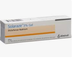
EL SOLARAZE, Diclofenac Gel (VOLTAREN)
Solaraze 3% Gel es un gel antiinflamatorio no esteroideo. Solaraze 3% Gel se aplica sobre la piel para tratar la queratosis actínica.
EDITORIAL ESPANOL:
====================
Hola amigos de la Cyber-Red, DERMAGIC, en esta ocasión les trae algunas referencias sobre un producto relativamente nuevo denominado SOLARAZE, compuesto por diclofenac sódico en gel al 3 % el cual ha sido puesto en venta en Alemania, Italia, Suiza, Canadá y probablemente otros países más. El producto es de la casa Hyal Pharmaceutical Corp. y tiene buen futuro, se está utilizando en lesiones precancerosas de la piel producidas por exposición al sol, fundamentalmente la queratosis actínica. También el producto parece ser beneficioso en las úlceras (aftosas) orales.
Aquí hay que hacer una aclaratoria: el GEL SOLARAZE es el MISMO VOLTAREN, y solo contiene diclofenac sódico al 3%. . El producto que contiene el diclofenac sódico al 3% mas ácido hialurónico su nombre comercial es ESQUERAT. Ambos se utilizan en las queratosis actínicas.
Hasta una próxima edición,,, saludos
Próximas ediciones: * LEISHMANIASIS CUTÁNEA, PENTAMIDINA E ITRACONAZOLE * ESPOROTRICOSIS, REVISIÓN
EDITORIAL ENGLISH:
===================
Hello friends of the Cyber-Net, DERMAGIC, in this occasion brings some references on a denominated relatively new product SOLARAZE, composed by sodium diclofenac in gel 3% and hyaluronic acid 2.5% which has been put in sale in Germany, Italy, Switzerland, France, Canada and probably other countries. The product is of The Hyal Pharmaceutical Corp.,and has good future. It is used in common precancerous skin lesion caused by overexposure to sunligh, mainly actinic keratosis. Also it seems to be that the product is beneficial in the oral aphthous ulcer.
Here we need to clarify: SOLARAZE GEL is the SAME VOLTAREN, and only contains 3% diclofenac sodium. The product that contains 3% diclofenac sodium plus hyaluronic acid is branded as ESQUERAT. Both are used for actinic keratoses.
Until a next edition, greetings
Next editions: * LEISHMANIASIS CUTANEOUS, PENTAMIDINE AND ITRACONAZOLE * SPOROTRICHOSIS UPDATE
=====================================================================
DERMAGIC/EXPRESS(18)
=====================================================================
E L SOLARAZE / T H E S O L A R A Z E =====================================================================
1.) An open study to assess the efficacy and safety of topical 3% diclofenac in a 2.5% hyaluronic acid gel for the treatment of actinic keratoses.
2.) Topical diclofenac/hyaluronic acid gel in the treatment of solar keratoses.
3.) The effect of hyaluronan on the in vitro deposition of diclofenac within the skin.
4.) The modulation of granulomatous tissue and tumour angiogenesis by diclofenac in
combination with hyaluronan (HYAL EX-0001).
5.) Angiostasis and vascular regression in chronic granulomatous inflammation induced by diclofenac in combination with hyaluronan in mice.
6.) The inhibition of colon-26 adenocarcinoma development and angiogenesis by topical
diclofenac in 2.5% hyaluronan.
7.) Sustained relief of oral aphthous ulcer pain from topical diclofenac in hyaluronan: a randomized, double-blind clinical trial.
8.) Hyaluronan as a drug delivery system for diclofenac: a hypothesis for mode of action.
9.) The depletion of substance P by diclofenac in the mouse.
10.) A controlled clinical investigation of 3% diclofenac/2.5% sodium hyaluronate topical gel in the treatment of uncontrolled pain in chronic oral NSAID users with osteoarthritis.
11.) Effect of vehicles on diclofenac permeation across excised rat skin.
12.) Solarase Approved In Canada For Precancerous Skin Lesions
13.) Solarase Approved In Europe For Treatment Of Sunspots
=======================================================================
1.) An open study to assess the efficacy and safety of topical 3% diclofenac in a 2.5% hyaluronic acid gel for the treatment of actinic keratoses.
=======================================================================
Author
Rivers JK; McLean DI
Address
Division of Dermatology, University of British Columbia and Vancouver Hospital and Health
Sciences Centre, Canada.
Source
Arch Dermatol, 133(10):1239-42 1997 Oct
Abstract
BACKGROUND: Actinic keratoses are potential precursors of invasive squamous cell
carcinoma; therefore, treatment is often recommended. Current topical treatments may cause considerable discomfort, pain, or skin irritation. This study was established to explore the
role, if any, of topical 3% diclofenac in 2.5% hyaluronic acid gel in the management of
actinic keratoses. OBSERVATIONS: An open-label study was conducted of topical 3%
diclofenac in 2.5% hyaluronic acid gel applied to 1 or more actinic keratoses. Patients
were instructed to apply 1.0 g of the gel twice daily for as many as 180 days. Treatment was
stopped earlier than 180 days if lesions were assessed as cleared. Twenty-nine adults were
treated for periods of 33 to 176 days (median, 62 days). Of the 29 subjects, 27 were
reevaluated 30 days after drug therapy discontinuation. Of the 27 patients, 22 (81%) had a
complete response and another 4 (15%) showed marked clinical improvement. The
preparation was generally well tolerated, although in 7 patients (24%) an irritant-type contact
dermatitis developed, which was confined to the treatment site. CONCLUSION: Topical
3% diclofenac in 2.5% hyaluronic acid gel may be a clinically useful topical agent for the
treatment of actinic keratoses.
=======================================================================
2.) Topical diclofenac/hyaluronic acid gel in the treatment of solar keratoses.
=======================================================================
Author
McEwan LE; Smith JG
Address
Peter MacCallum Cancer Institute, East Melbourne, Victoria, Australia.
Source
Australas J Dermatol, 38(4):187-9 1997 Nov
Abstract
A randomized double-blind controlled trial of 130 patients was performed to study the
efficacy and tolerability of topical 3% diclofenac in 2.5% hyaluronic acid (HA) gel
(active) versus gel containing 2.5% HA alone (control) in the treatment of solar keratoses.
Patients were asked to apply trial gel to the target lesion twice a day and also sunscreen once
a day for 24 weeks. The complete response rates were 29% for the active gel and 17% for
the control gel. The difference was not statistically significant (P = 0.14). A high percentage
of patients in both groups experienced a partial response to treatment (38% active, 45%
control) but there was no significant difference in the spectrum of response between the two
treatments (P = 0.18). Local adverse reactions occurred significantly more frequently in
patients using the active gel (29% compared to 5% using control gel, P = 0.0002).
=======================================================================
3.) The effect of hyaluronan on the in vitro deposition of diclofenac within the skin.
=======================================================================
Author
Brown MB; Marriott C; Martin GP
Address
Department of Pharmacy, King's College London, England.
Source
Int J Tissue React, 17(4):133-40 1995
Abstract
Hyaluronan is a glycosaminoglycan and has a protective, shock-absorbing and
structure-stabilizing role in connective tissue. Recently, it has been suggested that hyaluronan
can be used as a controlled and localized delivery vehicle for diclofenac in the treatment of a
variety of conditions including osteoarthritic pain, basal cell carcinoma and actinic keratosis.
The aim of this study was to examine the effects of hyaluronan on the in-vitro diffusion and
deposition of diclofenac within the skin. The studies involved full thickness, epidermal sheet
and stratum corneum human skin sections in an in-vitro Franz cell model, in which the
diffusion and deposition of 14C-labelled diclofenac and 3H-labelled hyaluronan was
investigated. The results showed that the diffusion of 14C-labelled diclofenac was sustained
and controlled by hyaluronan as compared to a buffer control, that a depot or reservoir of the
drug was formed in the epidermis and that it was probably this layer that determined the rate
of release of diclofenac within the skin. 3H-labelled hyaluronan was found to penetrate all
layers of the skin, although the rate of diffusion was much slower than that of labelled
diclofenac. Again, most of the activity was found to be retained within the epidermis, which
supports the hypothesis of the importance of this layer in the mode of action. The presence
within the epidermis of potential receptors for hyaluronan has recently been confirmed, and
this may partially explain the findings of the present in-vitro studies.
=======================================================================
4.) The modulation of granulomatous tissue and tumour angiogenesis by diclofenac in
combination with hyaluronan (HYAL EX-0001).
=======================================================================
Author
Freemantle C; Alam CA; Brown JR; Seed MP; Willoughby DA
Address
Department of Experimental Pathology, Saint Bartholomew's Hospital Medical College,
London, United Kingdom.
Source
Int J Tissue React, 17(4):157-66 1995
Abstract
In a novel application, hyaluronan has been utilized as a delivery system for topical and i.v.
therapeutics. Clinical trials and case reports show that topical diclofenac delivered in
hyaluronan (HYAL CT-1101) is effective against basal-cell carcinoma and actinic keratosis.
The effect of this drug formulation on tumour growth and angiogenesis, as well as
granulomatous tissue angiogenesis, has been investigated experimentally. The evidence that
hyaluronan has a permissive effect on the inhibition of granulomatous tissue angiogenesis by
diclofenac (as assessed by the carminel/gelatin vascular casting method) when injected into
the lesion or applied topically is reviewed. Topical diclofenac in hyaluronan also induces a
regression of the existing neo-vasculature of granulomatous tissue when applied
therapeutically. The diclofenac formulated in hyaluronan was also found to be profoundly
effective against the development of subcutaneous Colon-26 tumours in syngeneic balb/c
mice (T/C ratio after 12 days topical application of 0.174, p < 0.0001). Analysis of the
tumour vasculature showed that vascular development was retarded by 12 days. This was
shown by the reduction in the tumour density of carmine in the vascular casts, as well as
reduced blood-vessel density visualized by rat anti-mouse CD31 immunohistology.
Hyaluronan alone had a significant effect on tumour development with a 50% inhibition of
tumour growth and only a transient reduction in vascularity. The effects noted when
diclofenac is formulated in hyaluronan, and applied topically, could be related to
trans-dermal delivery and deposition properties of hyaluronan, and to the binding properties
of hyaluronan to areas of pathology with high expression of hyaluronan receptors such as
RHAMM, ICAM-1, and CD44.
=======================================================================
5.) Angiostasis and vascular regression in chronic granulomatous inflammation induced by diclofenac in combination with hyaluronan in mice.
=======================================================================
Author
Alam CA; Seed MP; Willoughby DA
Address
Department of Experimental Pathology, Saint Bartholomew's Hospital Medical College,
London, UK.
Source
J Pharm Pharmacol, 47(5):407-11 1995 May
Abstract
Angiostasis and vascular regression in chronic granulomatous inflammation was assessed in
mice induced with diclofenac in combination with hyaluronan. The local injection of 0.1 mL
HYAL EX-0001 (0.18% diclofenac in 2.5% hyaluronan) reduced granulomatous
development after six days treatment from 150.4 +/- 13.8 (0.18 saline) to 117.1 +/- 17.8 mg
(dry weight, n = 10), but not significantly when compared with 0.1 mL 2.5% hyaluronan
alone or diclofenac in 0.1 mL saline. Diclofenac administered in saline had no significant
effect when compared with saline control. The vascular density, expressed as carmine
content per mg dry weight tissue, in those animals treated with HYAL EX-0001 was also
significantly reduced to 5.27 +/- 0.55 micrograms mg-1 (P < 0.1, n = 10) when compared
with saline control (7.99 +/- 1.0), hyaluronan alone (7.20 +/- 1.0), and diclofenac in saline.
(7.36 +/- 1.28). A similar profile of activity was seen on topical application except that all
treatments did not affect granulomatous tissue development. On therapeutic dosing of mice
daily with HYAL EX-0001 from day 7 after induction of the granulomatous tissue, the
granulomatous tissue development was dramatically reduced from 111.67 +/- 4.40 mg (n =
14 on day 7) to 60.23 +/- 7.22 (P < 0.001, n = 8 on day 14) and 54.98 +/- 7.88 (P <
0.001, n = 8 on day 21). HYAL EX-0001 after 14 days of application significantly reduced
granulomatous tissue mass when compared with the hyaluronan-dosed control on day 21
(89.58 +/- 7.49, P = 0.01, n = 8).(ABSTRACT TRUNCATED AT 250 WORDS)
=======================================================================
6.) The inhibition of colon-26 adenocarcinoma development and angiogenesis by topical
diclofenac in 2.5% hyaluronan.
=======================================================================
Author
Seed MP; Brown JR; Freemantle CN; Papworth JL; Colville-Nash PR; Willis D; Somerville
KW; Asculai S; Willoughby DA
Address
Department of Experimental Pathology, Saint Bartholomew's Hospital and the Royal London
School of Medicine and Dentistry, United Kingdom. m.p.seed@mds.qmw.ac.uk
Source
Cancer Res, 57(9):1625-9 1997 May 1
Abstract
Topical diclofenac in 2.5% hyaluronan inhibits basal cell carcinoma, actinic keratosis, and
murine colon-26 growth in vivo. colon-26 tumor growth was preceded by angiogenesis and
reduced apoptotic and mitotic indices. Diclofenac reduced proliferation and viability in vitro,
and stimulated apoptosis. Hyaluronan inhibited proliferation and viability at 1 mg/ml but was
inactive below this level. Topical application of diclofenac inhibited tumor prostaglandin
synthesis and retarded angiogenesis and tumor growth (ratio of treatment:control, 0.174).
The mitotic index remained unaltered in vivo, whereas the apoptotic index and necrosis were
increased. Topical vehicle exhibited slight antitumor and antiangiogenesis activity. The
substantial quantities of diclofenac delivered locally in hyaluronan may exhibit antitumor
activity in similar fashion to those seen in vitro and explain its clinical efficacy.
=======================================================================
7.) Sustained relief of oral aphthous ulcer pain from topical diclofenac in hyaluronan: a randomized, double-blind clinical trial.
=======================================================================
Author
Saxen MA; Ambrosius WT; Rehemtula al-KF; Russell AL; Eckert GJ
Address
Department of Oral Surgery, Medicine and Pathology, Indiana University School of
Dentistry, Indianapolis, Ind., USA.
Source
Oral Surg Oral Med Oral Pathol Oral Radiol Endod, 84(4):356-61 1997 Oct
Abstract
OBJECTIVES: The purpose of this study was to test the hypothesis that topically applied
3% diclofenac in 2.5% hyaluronan reduces aphthous ulcer pain. STUDY DESIGN: A
randomized, double-blind, single dose study of 60 healthy adults with aphthous ulcers in three
treatment groups--3% diclofenac in 2.5% hyaluronan, 2.5% hyaluronan, 3% viscous
lidocaine--was undertaken. Visual analogue scale pain scores were obtained before and after
gel application and hourly, for up to 8 hours after gel application. Statistical analysis was
performed with repeated measures ANOVA with square root transformation and Bonferroni
correction. RESULTS: A 48% overall reduction in pain (p < 0.01) was observed 10 minutes
after gel application; however, no significant difference was found between the three topical
agents. A 35% to 52% pain reduction (p < 0.01) was reported 2 to 6 hours after the
application of diclofenac in hyaluronan, whereas hyaluronan gel alone and viscous lidocaine
failed to produce significant VAS reductions. CONCLUSIONS: A dose of 3% diclofenac
in 2.5% hyaluronan is an effective and novel treatment for this common, painful disorder.
=======================================================================
8.) Hyaluronan as a drug delivery system for diclofenac: a hypothesis for mode of action.
=======================================================================
Author
Moore AR; Willoughby DA
Address
Department of Experimental Pathology, St. Bartholomew's Hospital Medical College,
London, United Kingdom.
Source
Int J Tissue React, 17(4):153-6 1995
Abstract
Hyaluronan (HA) in combination with diclofenac is currently undergoing clinical trials as a
topical preparation in the management of osteoarthritic pain, basal-cell carcinoma and actinic
keratosis. These are clearly diverse pathologies, but in all cases substance P plays a central
role either directly or through secondary mediators such as prostaglandin E2 and nitric oxide.
A common mechanism for HA in combination with diclofenac in these conditions may be
through ameliorating the direct and indirect effects of substance P. Additionally, HA appears
to depot and hold diclofenac in the epidermis, thereby prolonging its pharmacokinetic
half-life. In rabbits, stenosis following balloon angioplasty is prevented by a subcutaneous
dose of HA, probably through blockade of cell-surface HA receptors (ICAM-1, CD44 &
RHAMM). The physicochemical properties of HA, and the binding of HA to HA receptors,
suggests that HA will have value as a novel drug delivery system.
=======================================================================
9.) The depletion of substance P by diclofenac in the mouse.
=======================================================================
Author
Papworth J; Colville-Nash P; Alam C; Seed M; Willoughby D
Address
Department of Experimental Pathology, William Harvey Research Institute, Saint
Bartholomew's and the Royal London Hospital School of Medicine and Dentistry, UK.
Source
Eur J Pharmacol, 325(2-3):R1-R2 1997 May 1
Abstract
Diclofenac in hyaluronan is analgesic and angiostatic. The depletion of substance P may be
a common mechanism. Mice received diclofenac, diclofenac in hyaluronan, or saline i.v.
for 5 days and snout substance P assessed: saline 2.80 +/- 0.23; 0.5 mg/kg diclofenac 2.03
+/- 0.20 (P < 0.05); diclofenac in hyaluronan 1.88 +/- 0.21 (P < 0.02); capsaicin 1.45 +/-
0.26 fmol/mg tissue (P < 0.005). Substance P recovered by 5 days (diclofenac in
hyaluronan, capsaicin) and 24 h (diclofenac). Diclofenac may deplete substance P in
analgesia, and hyaluronan prolong the depletion.
=======================================================================
10.) A controlled clinical investigation of 3% diclofenac/2.5% sodium hyaluronate topical gel in the treatment of uncontrolled pain in chronic oral NSAID users with osteoarthritis.
=======================================================================
Author
Roth SH
Address
Arthritis Center, Ltd., Phoenix, Arizona 85012, USA.
Source
Int J Tissue React, 17(4):129-32 1995
Abstract
Topical application, 4 times daily for 2 weeks, of the diclofenac-hyaluronan gel to 59
osteoarthritis patients induced a significantly greater (p = 0.057) analgesic effect than placebo
gel lacking the active substance applied to 60 control patients.
=======================================================================
11.) Effect of vehicles on diclofenac permeation across excised rat skin.
=======================================================================
Author
Takahashi K; Suzuki T; Sakano H; Mizuno N
Address
Faculty of Pharmaceutical Sciences, Mukogawa Women's University, Hyogo, Japan.
Source
Biol Pharm Bull, 18(4):571-5 1995 Apr
Abstract
The in vitro percutaneous permeation of diclofenac from various vehicles was examined
using rat abdominal skin as a model membrane. The oleaginous vehicles used in this study
consisted of three components: i.e. fatty acid, fatty acid ester and nonpolar oil. The
solubilities of sodium diclofenac in formulated vehicles were above 0.2 M. The effect of
each oleaginous component in the vehicle on the permeation of diclofenac across the skin
was in the following order: oleic acid > isostearic acid, diisopropyl adipate = diethyl
sebacate > Panasate 875 and squalane > liquid paraffin. To clarify the reason for the
differences in permeation among the fatty acid esters, the release of diclofenac through
either porous or lipoidal membranes from these vehicles in vitro and the solubility of sodium
diclofenac in the vehicles were studied. However, no relationship was observed between
the release rate or solubility and skin permeability. The skin permeation of diclofenac
increased following pretreatment with diisopropyl adipate or diethyl sebacate, but not with
middle chain triglyceride (Panasate 875). These results suggested that the main reason may
be the enhancement effect of fatty acid esters. Emulsions and creams containing 3% sodium
diclofenac were prepared using the above oleaginous vehicles. A large flux and short lag
time were observed in these preparations compared with an aqueous suspension of sodium
diclofenac. The incorporation of urea significantly enhanced the permeation of diclofenac
from these preparations. These results suggest that the emulsion and cream prepared in this
study are useful for development for practical purposes.
=======================================================================
12.) Solarase Approved In Canada For Precancerous Skin Lesions
=======================================================================
MISSISSAUGA, ON -- May 20, 1998 -- The Therapeutic Products
Directorate of Health Canada (formerly HPB) has granted marketing approval
to Hyal Pharmaceutical Corp.’s Solarase™, a topical gel formulation to treat
actinic keratosis (AK), precancerous skin lesions caused by exposure to the
sun.
AK is a common precancerous skin lesion caused by overexposure to sunlight.
A small number of these lesions will develop into squamus cell carcinoma, a
malignant skin disease. According to The Skin Cancer Foundation of the
United States, AK affects one in six people world-wide.
Hyal expects to file a submission to the FDA in the third quarter seeking
approval to market Solarase in the U.S.
Solarase utilises Hyal's patented HIT technology which targets and retains
drugs in areas of dermal pathology. The benefits to the patient are minimal
systemic drug absorption and outstanding safety.
=======================================================================
13.) Solarase Approved In Europe For Treatment Of Sunspots
=======================================================================
MISSISSAUGA, ON -- May 4, 1998 -- Hyal Pharmaceutical Corp.’s lead
product Solarase, a topical therapy for the treatment of actinic keratosis or
sunspots, has received regulatory approval for marketing in Germany, Sweden,
Italy and France.
Actinic keratosis is a sun exposure-related precancerous skin condition
affecting one in six adults worldwide and is the forerunner of one of the most
common forms of skin cancer -- squamous cell carcinoma..
Compared to other actinic keratosis treatments, Solarase is non-invasive,
non-scarring and extremely well tolerated by patients.
======================================================================
DATA-MÉDICOS/DERMAGIC-EXPRESS No (18) 20/11/98 DR. JOSE LAPENTA R. DERMATÓLOGO
======================================================================
Produced by Dr. José Lapenta R. Dermatologist Venezuela 1.998-2.0024
Producido por Dr. José Lapenta R. Dermatólogo Venezuela 1.998-2.024
Tlf: 0414-2976087 - 04127766810


.png)